Ori Shokek, M.D., Moody D. Wharam, Jr., M.D., F.A.C.R.

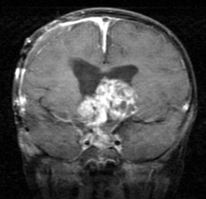
Craniopharyngioma is a brain tumor that occurs in children. It is seen in the suprasellar region of the brain, a centrally located area adjacent to critical structures including the nerves that conduct visual signal from the eyes to the brain, the ventricular system which governs the flow and pressure of the fluid that surrounds the brain, and the pituitary gland and hypothalamus which produce important regulatory hormones. Because the tumor does not invade and destroy the normal tissues that surround it, and as it does not spread distantly away from its local origin, it is said to be a benign tumor. However, as it grows, the tumor can damage those critical structures that surround it, resulting in disturbance of their normal function. Such disturbance, commonly in vision, brain pressure, and hormone production, is typically what first brings the patient to medical attention.
Craniopharyngioma is curable in the majority of patients. Treatment for this tumor most often involves surgery and radiation therapy. Although complete surgical resection can be sufficient for cure, the tumor is often simply sticky, and there can be significant difficulty in removing it from the critical structures to which it may adhere. The attempt at complete surgical resection may result in damage to those same critical structures. The same neurologic disturbances that can be caused by the tumor itself can also result from such surgery, and they can be life-long. In addition, MRI or CT scans performed after an attempt at a complete resection sometimes reveal residual tumor. Therefore, an established treatment approach in many medical centers in the United States involves a partial resection of the tumor (often termed decompressive surgery) followed by radiation therapy. As far as cure, results with partial resection and radiation therapy are often as good as complete surgical resection.

Radiation therapy has side effects. There can be hormonal disturbances, although those caused by radiation therapy are typically easier to manage. Visual deficits are uncommon. Additional side effects exist but depend on the particular patient, on his or her age, and on the extent and size of the tumor.
It is important for patients and families to be familiar with the types of treatment their physicians might recommend. Most people have a basic understanding of what surgery is, but not everyone is familiar with radiation therapy. Radiation therapy typically uses X-rays, similar to those used by a CT scan. ‘External beam’ radiation therapy is the most common way treatment is given. Here, radiation is directed toward the patient from an external machine, usually a linear accelerator. Multiple beams are used, all individually shaped and all converging upon the tumor, such that the tumor receives the sum of their individual doses of radiation; the surrounding brain structures receive only the contribution of each individual beam. Treatment is typically given in daily sessions over a course of approximately six weeks, with a small fraction of the total dose delivered on any given day. Larger doses of radiation given over a shorter time course have the potential to cause greater side effects. However, certain small craniopharyngiomas are amenable to even one single large dose of radiation, if sparing of surrounding critical structures is possible. For such selected tumors, the Gamma Knife is a highly specialized external beam machine which is quite effective.
X-rays are not the only type of radiation used in the treatment of craniopharyngioma. Protons are another type and have the advantage that their dose can be directed to a particular depth as it is aimed at the tumor. Lastly, some craniopharyngiomas which have fluid filled cysts are amenable to brachytherapy. This is a way of delivering radiation to the tumor by inserting a needle into it and injecting a radioactive liquid that emits radiation. As brachytherapy allows radiation to be delivered from the inside of the tumor, it has the advantage that only negligible doses of radiation reach adjacent normal brain structures.
In summary, craniopharyngioma is a benign tumor seen in children. Its cure rate is high, and treatment is individualized and commonly includes surgery and radiation therapy. Potential side effects are carefully considered when choosing the treatment approach. Physicians at medical centers dedicated to the management of childhood brain tumors are able to offer the appropriate level of expertise and can optimally tailor treatment to the individual patient.
Moody D. Wharam, M.D., F.A.C.R. was Professor of Radiation Oncology in the Department of Radiation Oncology and Molecular Radiation Sciences at the Johns Hopkins University School of Medicine, Baltimore, Maryland. He died August 10, 2018.
Ori Shokek, M.D. was Dr. Wharum’s colleague at Johns Hopkins, Chief Resident in Radiation Oncology. Dr. Ori Shokek is now at WellSpan Radiation Oncology. There are two locations: York, PA and Baltimore, Maryland. Drs. Wharam and Shokek co-authored this article.
This article will be reviewed for any updates shortly.
